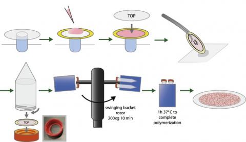
Congratulations to Laralynne Przybyla and coauthors on their publication in Methods!
Abstract: The way cells are organized within a tissue dictates how they sense and respond to extracellular signals, as cues are received and interpreted based on expression and organization of receptors, downstream signaling proteins, and transcription factors. Part of this microenvironmental context is the result of forces acting on the cell, including forces from other cells or from the cellular substrate or basement membrane. However, measuring forces exerted on and by cells is difficult, particularly in an in vivo context, and interpreting how forces affect downstream cellular processes poses an even greater challenge. Here, we present a simple method for monitoring and analyzing forces generated from cell collectives. We demonstrate the ability to generate traction force data from human embryonic stem cells grown in large organized epithelial sheets to determine the magnitude and organization of cell–ECM and cell–cell forces within a self-renewing colony. We show that this method can be used to measure forces in a dynamic hESC system and demonstrate the ability to map intracolony protein localization to force organization.

